Upper Thigh Muscles Anatomy - Ch. 10 / 11 Muscle / Tissue - Anatomy & Physiology 1 with ... - Muscles of the upper and lower leg.
Upper Thigh Muscles Anatomy - Ch. 10 / 11 Muscle / Tissue - Anatomy & Physiology 1 with ... - Muscles of the upper and lower leg.. 430) is a flat, quadrangular muscle, situated at the anterior part of the upper and medial aspect of the thigh. Leg muscle anatomy for figurative artists. 2, vastus medialis & intermedius muscles. Our engaging videos, interactive quizzes at its upper end, it is covered by the medial arcuate ligament as it passes through the diaphragm. The thigh is the area between the hip and the knee joint.
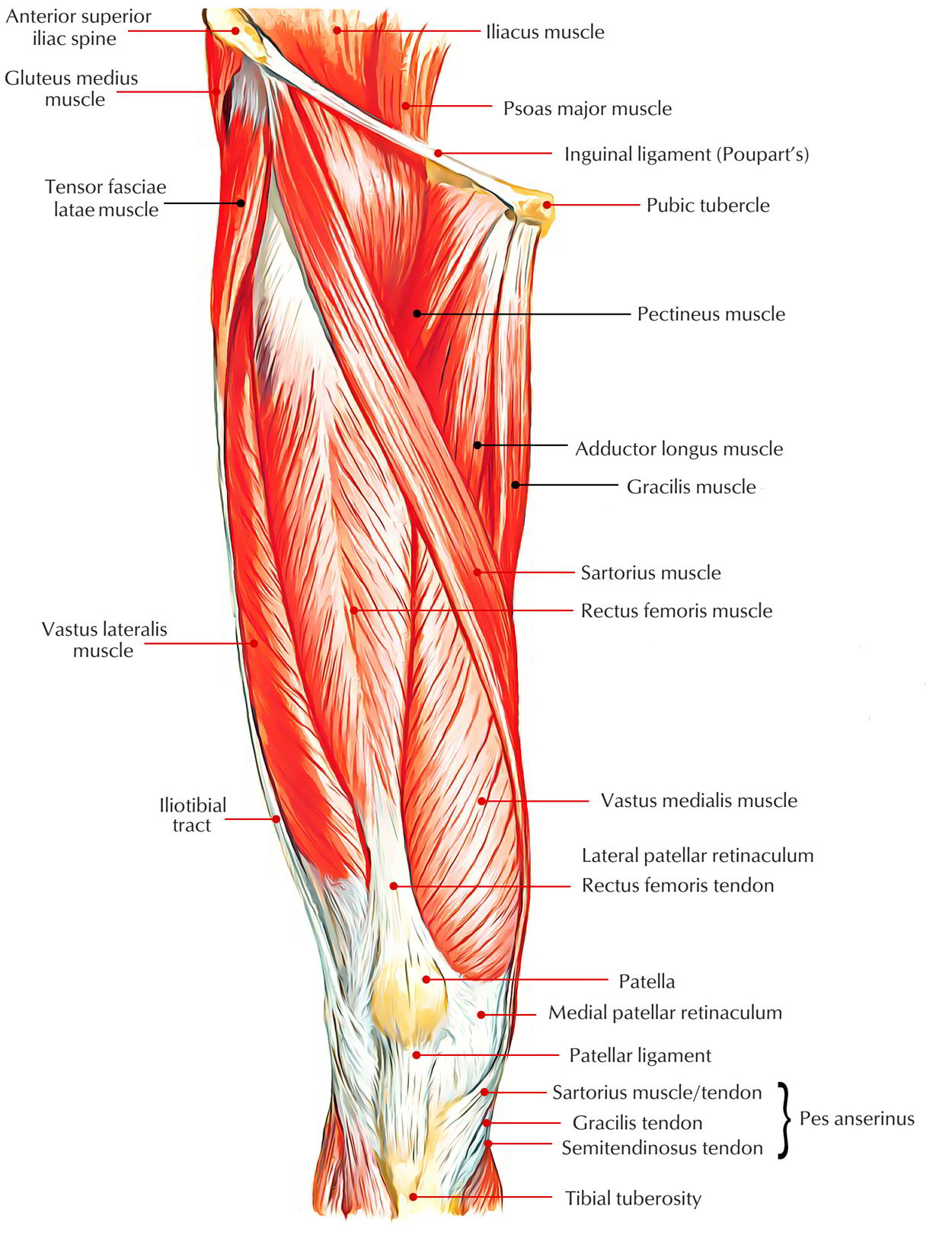
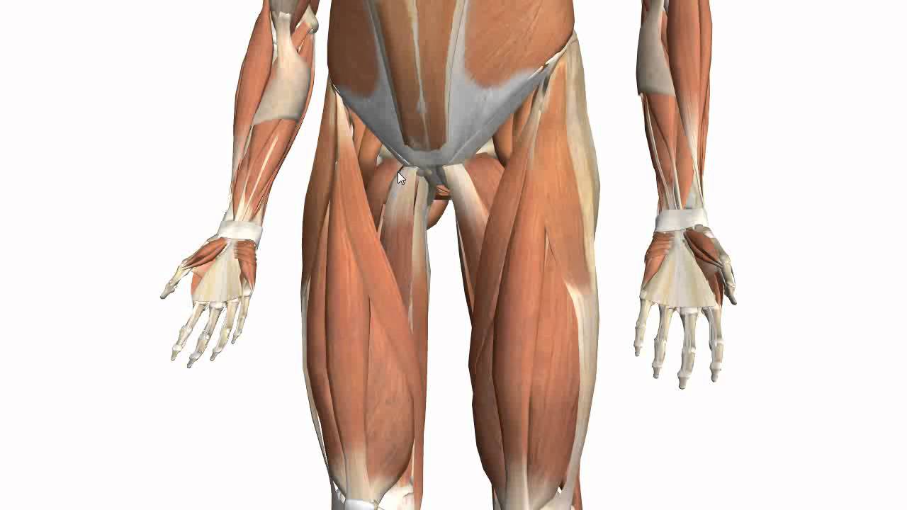
The single bone in the thigh region is called the femur. Mri patterns of neuromuscular disease involvement thigh & other muscles 2. Microscopic anatomy of skeletal muscle. Like the forearm, the upper leg, or thigh, has a dense arrangement of many muscles. The sartorius muscle attaches to the hip bone (iliac spine), travels down the front of the thigh moving toward the inside of the thigh, and connects to the inside of the shin bone (tibia).
Easy Notes On 【Muscles of Anterior Compartment of The ... from www.earthslab.com The single bone in the thigh is called the femur. This webpage presents the anatomical structures found on thigh mri. In human anatomy, the thigh is the area between the hip (pelvis) and the knee. Muscles of the arm and forearm diagram. The hamstrings flex the knee joint and extend the thigh to the back side of the body. 430) is a flat, quadrangular muscle, situated at the anterior part of the upper and medial aspect of the thigh. At the top, there is the pelvis bones which do not belong to the lower limb anatomy, but are part of the the outer head of this muscle originates from the upper edge of the thigh bone and goes downward to the knee cap. The extrinsic group originate from the torso and attach to the bones of the both groups are innervated by the ulnar and median nerve.
Related posts of muscle anatomy of upper thigh shoulder muscles anatomy.
The thigh is the area between the hip and the knee joint. A complete list of muscular system quizzes; This webpage presents the anatomical structures found on thigh mri. It is a powerful extensor of the thigh. Anatomynote.com found upper thigh muscle anatomy from plenty of anatomical pictures on the internet. Popular study materials from anatomy and cell biology 306. Want to learn more about it? In clinical anatomy the thigh muscles are divided into three groups: Anatomy and function of forearm muscles. We look at the associated symptoms and treatment options. It inserts onto the linea aspera of the femur. These muscles are extremely important for knee extension and trunk or hip flexion. 430) is a flat, quadrangular muscle, situated at the anterior part of the upper and medial aspect of the thigh.
Microscopic anatomy of skeletal muscle. It contains both an anterior and posterior compartment, and each is further divided into layers. Let's begin with the skeletal anatomy. Like the forearm, the upper leg, or thigh, has a dense arrangement of many muscles. Small and deep muscles which mainly externally rotate the thigh at the hip joint and stabilize the pelvis.
95 best Drawing: Anatomy images on Pinterest | Human ... from i.pinimg.com Pain in the upper thigh can be difficult to diagnose because this area of the body contains many muscles, tendons, and ligaments. Anatomynote.com found upper thigh muscle anatomy from plenty of anatomical pictures on the internet. Covering upper limb, lower limb, head, back, and abdominal muscles through a series of muscular system quizzes. They are used in walking, running, and many other physical activities. In this section, learn more about the anatomy of the muscles of the upper limb… Like the forearm, the upper leg, or thigh, has a dense arrangement of many muscles. It inserts onto the linea aspera of the femur. The thigh is the area between the hip and the knee joint.
We look at the associated symptoms and treatment options.
Muscles and ligaments work together to support the spine, hold it upright, and control movement during rest and activity. The pectineus is a flat, quadrangular muscle situated at the anterior part of the upper and medial aspect of the thigh. Muscles of the arm and forearm diagram. It arises from the pectineal line, and to a slight. They are further categorized according function such as flexion, extension, or rotation. 2, vastus medialis & intermedius muscles. Small and deep muscles which mainly externally rotate the thigh at the hip joint and stabilize the pelvis. Involved early gray = muscle: #1 free online anatomy resource. Anatomy and function of forearm muscles. Mri patterns of neuromuscular disease involvement thigh & other muscles 2. At the top, there is the pelvis bones which do not belong to the lower limb anatomy, but are part of the the outer head of this muscle originates from the upper edge of the thigh bone and goes downward to the knee cap. Muscles are named according to their shape, location, or a combination.
You can click the image to magnify if you cannot see clearly. Quadriceps muscle is made of rectus femoris, vastus lateralis, vastus medialis and vastus intermedius. 430) is a flat, quadrangular muscle, situated at the anterior part of the upper and medial aspect of the thigh. Learn about the anatomy and purpose of the hamstrings, a group of muscles at the rear of the upper leg, plus get info on exercises and stretches. The extrinsic group originate from the torso and attach to the bones of the both groups are innervated by the ulnar and median nerve.
Muscles of the Thigh Part 2 - Medial Compartment - Anatomy ... from i.ytimg.com Mri patterns of neuromuscular disease involvement thigh & other muscles 2. A muscle of the medial thigh that originates on the pubis. In this section, learn more about the anatomy of the muscles of the upper limb… Anatomy and function of forearm muscles. Involved early gray = muscle: Muscles and ligaments work together to support the spine, hold it upright, and control movement during rest and activity. Body muscle chart muscle diagram body anatomy organs human body anatomy upper limb anatomy anatomy bones. 2, vastus medialis & intermedius muscles.
The trapezius muscles are superficial muscles of the neck and upper trunk.
It arises from the pectineal line, and to a slight. When this muscle contracts, it. Quadriceps muscle is made of rectus femoris, vastus lateralis, vastus medialis and vastus intermedius. Anatomynote.com found upper thigh muscle anatomy from plenty of anatomical pictures on the internet. Taken together they form a diamond shape. Anatomy and function of forearm muscles. It is part of the lower limb. Anatomically, it is part of the lower limb. A complete list of muscular system quizzes; They are used in walking, running, and many other physical activities. Muscles of the upper and lower leg. The upper limb muscles fall into three groups. This webpage presents the anatomical structures found on thigh mri.
They are further categorized according function such as flexion, extension, or rotation upper thigh anatomy. Anatomynote.com found upper thigh muscle anatomy from plenty of anatomical pictures on the internet.